


Good morning,
Today, I wish to share with the readers, particularly the ENT fraternity, a refresher on what to observe when reviewing a CT paranasal sinus image. These are my undergraduate notes, so they cover the basics (credit to Prof. Dr. Salina Husain). I hope this entry proves helpful to you surgeons in some manner.
MNEMONIC: CLOSSEF
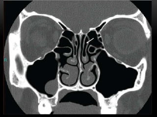
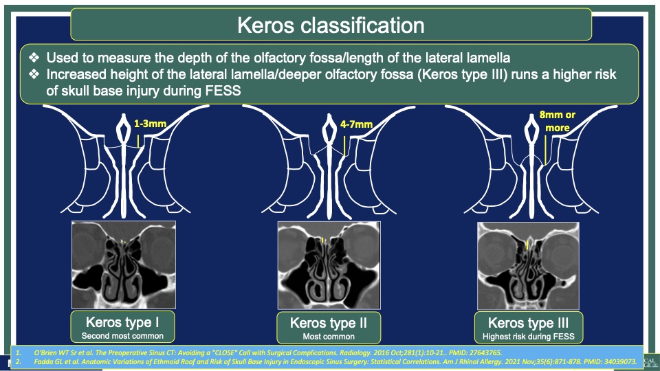
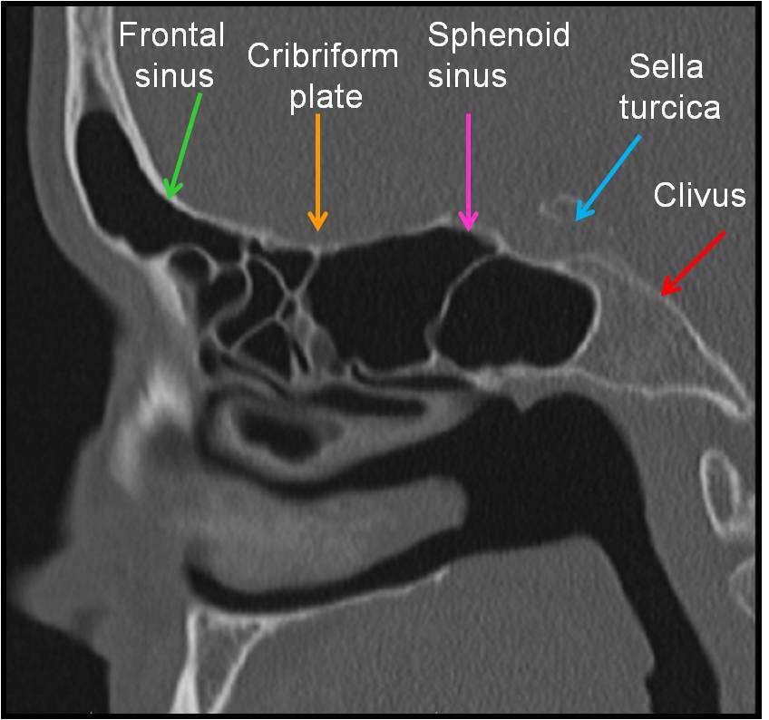
C – Cribiform plate (keros classification) – to assess the depth of olfactory fossa, length of lateral lamella, symmetry, slope or any dehiscence.
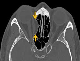
L – Lamina Papyracea – look for dehiscence, so if there is dehiscence make sure to be extra careful when using microdebrider around this area as incident such as injuring the orbital fat or rectus muscle could occur.
O – Onodi cell – if present, to assess its relationship to optic nerve, ICA, dehiscence (coronal and axial) and presence of horizontal septa.

S – Sphenoid sinus – pneumatization, septations, dehiscence of carotid artery or optic nerve.
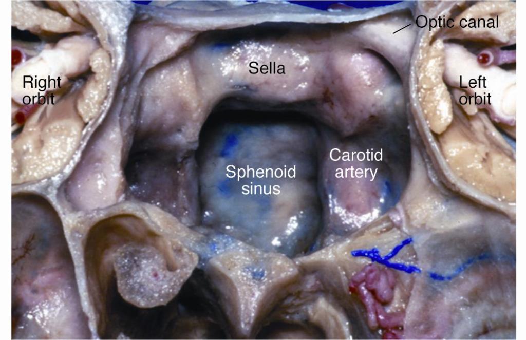
S – Skull base – dehiscence, slope (examine sagittal plane of CT scan)
E – Ethmoidal artery position (skull base or pedicle), symmetrical? Usually anterior ethmoidal artery (~40% hanging )
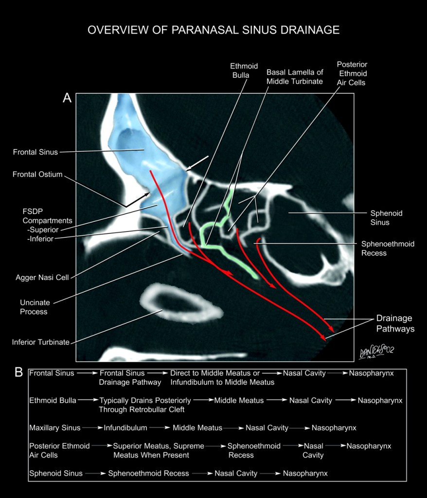
FSDP – frontal sinus drainage pathway – presence or absence and degree of penumatization of various cells and lamellas – ethmoid bulla, supra-bullar, fronto-bullar, supra-orbital, fronto-ethmoidal cells, and the Agger Nasi Cell.
If you find this brief entry helpful, please consider sharing this knowledge with others.


Leave a comment